Tuesday, August 03, 2004
THE NEVERENDING TRAUMA WORKUP...
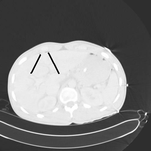
20-ish y/o restrained driver in SVMVC. +LOC with complaints of abdominal pain and facial pain. Hemodynamics are stable. Otherwise healthy. Undergoes CT of abdomen:

The black lines point to two small areas of free air. He is also found to have a nasal bone fracture but no other acute findings on CT. He is taken to the OR for an exp lap with findings of a jejunal tear which is repaired without difficulty. Two days later he complains of inability to move his right leg. Sensation to coarse touch and proprioception is intact. He has diminished pulses in that foot. Some pain on passive range of motion but no bony crepitus. A noninvasive vascular study is ordered and reveals an ankle-brachial index of 0.9 on the left and 0.6 on the right. An angiogram is ordered:
This is a view of his superficial femoral artery. Looks good but you can see some problems beginning distally.
What you see here is occlusion of his popliteal artery above the knee with reconstitution of the below-knee popliteal and filling of the trifurcation. Notice the size of the collaterals.

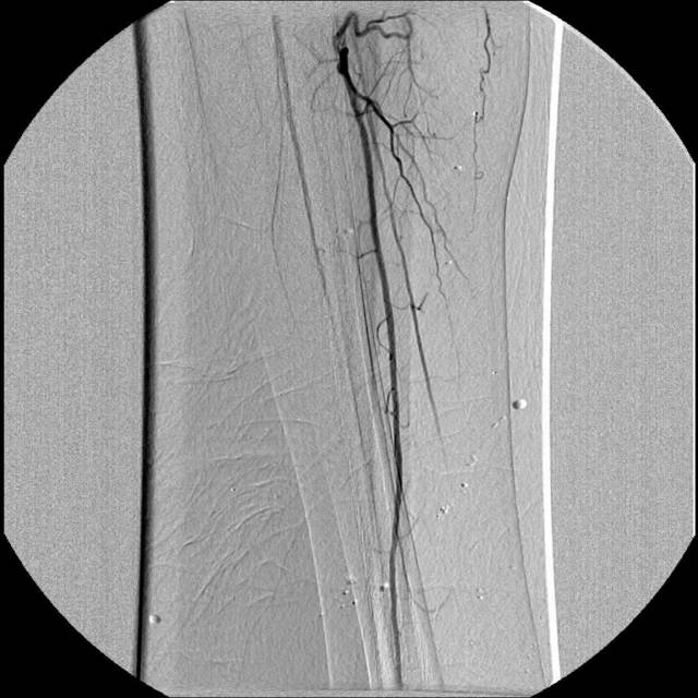
Unfortunately he appears to only have one-vessel runoff to his foot. Our diagnosis at this time was popliteal artery injury, likely due to a knee dislocation which had spontaneously reduced. We were not totally convinced due to his lack of joint instability. The initial plan was to attempt a stent-graft across the area of injury. (By this time my vascular surgeon partner was along for the ride.) We were able to get a wire and catheter through a collateral but not into the main artery. After further angiography we determined that what the patient had was not an acute injury but popliteal artery entrapment syndrome. The patient then underwent a popliteal to popliteal bypass with reversed saphenous vein. Upon further questioning he did state he had experienced worsening leg pain over the past few weeks but had attributed it to "soreness". This patient fits the profile in the article very well. Young, active and otherwise healthy. It was fortunate the condition was discovered while his foot was still viable, since embolization can be a complication. The size of the collateral vessels on his angiogram provide an indication of the long-standing nature of this problem when compared to a more acute change.

Where the vessels are smaller. He is doing well and has regained motor function in his lower extremity |
20-ish y/o restrained driver in SVMVC. +LOC with complaints of abdominal pain and facial pain. Hemodynamics are stable. Otherwise healthy. Undergoes CT of abdomen:
The black lines point to two small areas of free air. He is also found to have a nasal bone fracture but no other acute findings on CT. He is taken to the OR for an exp lap with findings of a jejunal tear which is repaired without difficulty. Two days later he complains of inability to move his right leg. Sensation to coarse touch and proprioception is intact. He has diminished pulses in that foot. Some pain on passive range of motion but no bony crepitus. A noninvasive vascular study is ordered and reveals an ankle-brachial index of 0.9 on the left and 0.6 on the right. An angiogram is ordered:

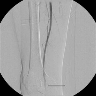
This is a view of his superficial femoral artery. Looks good but you can see some problems beginning distally.

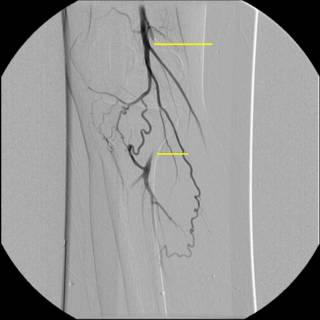
What you see here is occlusion of his popliteal artery above the knee with reconstitution of the below-knee popliteal and filling of the trifurcation. Notice the size of the collaterals.
Unfortunately he appears to only have one-vessel runoff to his foot. Our diagnosis at this time was popliteal artery injury, likely due to a knee dislocation which had spontaneously reduced. We were not totally convinced due to his lack of joint instability. The initial plan was to attempt a stent-graft across the area of injury. (By this time my vascular surgeon partner was along for the ride.) We were able to get a wire and catheter through a collateral but not into the main artery. After further angiography we determined that what the patient had was not an acute injury but popliteal artery entrapment syndrome. The patient then underwent a popliteal to popliteal bypass with reversed saphenous vein. Upon further questioning he did state he had experienced worsening leg pain over the past few weeks but had attributed it to "soreness". This patient fits the profile in the article very well. Young, active and otherwise healthy. It was fortunate the condition was discovered while his foot was still viable, since embolization can be a complication. The size of the collateral vessels on his angiogram provide an indication of the long-standing nature of this problem when compared to a more acute change.
Where the vessels are smaller. He is doing well and has regained motor function in his lower extremity |
![]()


